$479.00 Original price was: $479.00.$114.00Current price is: $114.00.
 Purchase this course you will earn 114 Points worth of $11.40
Elevate your skills with the CPD – MS068 – Abdominal Radiology Mini Series course, available for just $479.00 Original price was: $479.00.$114.00Current price is: $114.00. on Utralist.com! Browse our curated selection of over 60,000 downloadable digital courses across diverse Everything Else. Benefit from expert-led, self-paced instruction and save over 80%. Start learning smarter today!
Purchase CPD – MS068 – Abdominal Radiology Mini Series courses at here with PRICE $479 $114
MS068 – Abdominal Radiology Mini Series
£347.00 (+VAT)
12 months access to recordings and course materials is included. Please note that these are webinar recordings and not live events. Full details on how to access the Mini Series will be emailed to you.
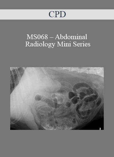
Accurate evaluation of abdominal radiographs can be challenging, as the majority of abdominal organs are of soft tissue opacity and, when superimposed on an abdominal radiograph, can be difficult to distinguish from each other. Consistent interpretation of abdominal radiographs requires a logical approach, as well as a good knowledge of normal radiographic anatomy, enabling you to recognise the presence and location of any abnormalities. During this Mini Series, we will work our way logically through the abdominal cavity of the dog and cat, reviewing the normal radiographic appearance of the abdominal structures and using that knowledge to identify and evaluate radiographic abnormalities commonly encountered in abdominal disease. At the end of each webinar, a multiple-choice film-reading quiz will provide an opportunity to put the theory into practice.
- Join RCVS & European Specialist in Veterinary Diagnostic Imaging Esther Barrett for three 2-hour sessions
- Get all the help you need with your Abdominal Radiology in practice
- Learn about recent advances that you can apply to your patients
- Lots of case-based discussion and videos to illustrate key points
- Comprehensive notes to download
- Self-assessment quiz to ‘release’ your 8 hours CPD certification (don’t worry, you can take them more than once if you don’t quite hit the mark first time)
- A whole year’s access to recorded sessions for reviewing key points
- Superb value for money – learn without travelling
- This Mini Series was originally broadcast in August 2014
Programme
Session 1
Getting the Best out of Your Abdominal Radiographs and Evaluating the Abdominal Space, Liver and Spleen
- Obtaining diagnostic abdominal radiographs – what are the potential challenges?
- Principles of interpretation – remember Roentgen signs?
- Abdominal radiology vs ultrasound – do I need both?
- A logical approach to abdominal masses
- Evaluating the abdominal space
- Normal radiographic anatomy of the peritoneal and retroperitoneal spaces
- Identifying free fluid and free gas
- Typical radiographic features of peritonitis
- Typical radiographic features of abdominal lymphadenopathy
- Evaluating the liver
- Recognising changes in hepatic size, shape and opacity
- Typical radiographic features of liver masses, metabolic hepatopathy, congenital porto-systemic shunts and chronic liver disease
- Evaluating the spleen
- Recognising changes in splenic size, shape and opacity
- Typical radiographic features of splenic masses and splenic torsion
- What could ultrasound add to the evaluation of the abdominal space, liver and spleen?
- Test yourself: Examples of commonly encountered hepatic, splenic and abdominal space disease
Evaluating the Urogenital Tract
- Evaluating the kidneys, ureters, bladder and urethra
- Recognising changes in renal size, shape and opacity
- Urinary tract contrast studies
- Radiographic features of renal & ureteral calculi, nephritis / pyelonephritis, hydronephrosis, ectopic ureters, renal neoplasia, renal cysts and renal and/or ureteral rupture
- Radiographic features of cystitis, bladder and urethral calculi, bladder and urethral neoplasia, bladder and urethral rupture and bladder diverticula
- Evaluating the ovaries, uterus and prostate
- Radiographic features of pregnancy, pyometra, uterine and ovarian neoplasia
- Radiographic features of prostatic disease, paraprostatic cysts and cryptorchidism
- What could ultrasound add to the evaluation of the uro-genital tract?
- Test yourself: Examples of commonly encountered hepatic, splenic and abdominal space disease
- Test yourself: Examples of commonly encountered uro-genital abnormalities
Evaluating the Gastro-intestinal Tract
- Evaluating the stomach
- Normal radiographic anatomy and variations
- Identifying gastric foreign bodies
- Typical radiographic features of gastric dilation and volvulus
- Evaluating the small intestine
- Normal radiographic anatomy
- Logical evaluation of changes in the size, shape, opacity and position if the small intestinal loops
- Evaluating the pancreas
- Can I recognise pancreatitis from an abdominal radiograph?
- Evaluating the large intestine
- Normal radiographic anatomy
This webinar mini-series is suitable for all vets in practice, and will be of particular interest to those wishing to improve their confidence in the interpretation of abdominal radiographs.
Course Feedback:
“This course will help me in practice a great deal – improved confidence in interpretation, more likely to pick up on abnormalities that would previously overlooked”
“I thought it was very thorough, with good explanations. It will help me to identify abnormalities on abdominal radiography”
“The recordings were particularly useful as I could pause and study the radiographs. I feel much more confident in my interpretive skills”
Purchase CPD – MS068 – Abdominal Radiology Mini Series courses at here with PRICE $479 $114
Cultivate continuous growth with the CPD – MS068 – Abdominal Radiology Mini Series course at Utralist.com! Unlock lifetime access to premium digital content, meticulously designed for both career advancement and personal enrichment.
- Lifetime Access: Enjoy limitless access to your purchased courses.
- Exceptional Value: Benefit from savings up to 80% on high-quality courses.
- Secure Transactions: Your payments are always safe and protected.
- Practical Application: Gain real-world skills applicable to your goals.
- Instant Accessibility: Begin your learning journey immediately after buying.
- Device Compatible: Access your courses seamlessly on any device.
Transform your potential with Utralist.com!
Related products
Everything Else
= 47 Points
Everything Else
Brian Tracy – 21st Century Sales Training for Elite Performance
= 47 Points
Everything Else
= 81 Points
Everything Else
= 43 Points
Everything Else
= 56 Points
Everything Else
= 128 Points
Everything Else
= 89 Points
Everything Else
= 85 Points
Login

 Purchase this course you will earn 114 Points worth of $11.40


